World · India Bureau
Scientists develop dye-free method to detect immune cell activation
Researchers have developed a breakthrough technique that can identify activated immune cells with 94% accuracy in just two hours without requiring chemical dyes. The innovation analyzes natural light emissions from immune cell molecules to distinguish between active and dormant states.
LSN India ·
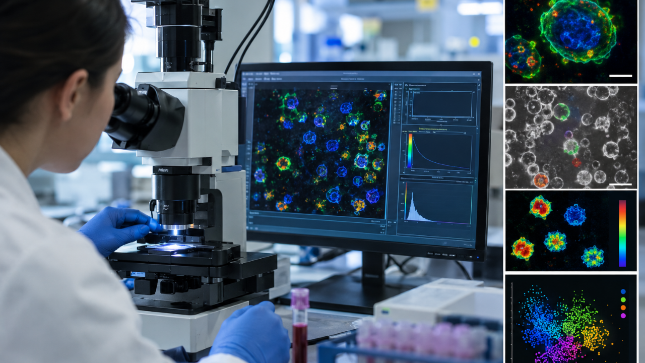
A new scientific method has emerged that promises to revolutionize the study of immune cell function by eliminating the need for chemical dyes in cellular imaging. The technique works by analyzing light naturally emitted from molecules within immune cells, enabling researchers to accurately determine whether cells are in an activated or resting state.
The innovation achieves a 94% accuracy rate in identifying immune cell activation status within a two-hour timeframe. By examining cellular metabolism at the single-cell level without introducing external dyes, the method provides a cleaner, more direct approach to understanding immune cell behavior and function.
Traditional methods of immune cell analysis have relied on fluorescent dyes and stains to visualize cellular states, which can sometimes interfere with the cells themselves or complicate the imaging process. This dye-free approach offers a more natural window into cellular activity, potentially providing researchers with more accurate biological data.
The breakthrough could have significant implications for immunology research and clinical diagnostics. By streamlining the detection process and improving accuracy, the technique may accelerate the pace of immune-related research and enhance our understanding of how immune cells respond to infections, vaccines, and other health challenges.
The development reflects growing interest in non-invasive, label-free methods for cellular analysis that can improve both the speed and reliability of biological research across the region and globally.